Dorsalis pedis artery
| Dorsalis pedis artery | |
|---|---|
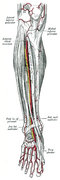 Anterior tibial artery, dorsalis pedis artery and the muscles and bones of the leg (anterior view). | |
| Details | |
| Source | anterior tibial artery |
| Branches | First dorsal metatarsal artery and Deep plantar artery |
| Supplies | dorsal surface of the foot |
| Identifiers | |
| Latin | arteria dorsalis pedis |
| TA | A12.2.16.048 |
| FMA | 43915 |
Anatomical terminology [edit on Wikidata] | |
In human anatomy, the dorsalis pedis artery (dorsal artery of foot), is a blood vessel of the lower limb that carries oxygenated blood to the dorsal surface of the foot. It is located 1/3 from medial malleolus. It arises at the anterior aspect of the ankle joint and is a continuation of the anterior tibial artery. It terminates at the proximal part of the first intermetatarsal space, where it divides into two branches, the first dorsal metatarsal artery and the deep plantar artery. The dorsalis pedis communicates with the plantar blood supply of the foot through the deep plantar artery.
Along its course, it is accompanied by a deep vein, the dorsalis pedis vein.
Palpation of the dorsalis pedis artery pulse
The dorsalis pedis artery pulse can be palpated readily lateral to the extensor hallucis longus tendon (or medially to the extensor digitorum longus tendon) on the dorsal surface of the foot, distal to the dorsal most prominence of the navicular bone which serves as a reliable landmark for palpation.[1] It is often examined, by physicians, when assessing whether a given patient has peripheral vascular disease. It is absent, unilaterally or bilaterally, in 2–3% of young healthy individuals.[2]
References
^ Mowlavi, A; Whiteman, J; Wilhelmi, BJ; Neumeister, MW; McLafferty, R (2002). "Dorsalis pedis arterial pulse: palpation using a bony landmark". Postgraduate Medical Journal. 78 (926): 746–7. doi:10.1136/pmj.78.926.746. PMC 1757948. PMID 12509693..mw-parser-output cite.citation{font-style:inherit}.mw-parser-output .citation q{quotes:"""""""'""'"}.mw-parser-output .citation .cs1-lock-free a{background:url("//upload.wikimedia.org/wikipedia/commons/thumb/6/65/Lock-green.svg/9px-Lock-green.svg.png")no-repeat;background-position:right .1em center}.mw-parser-output .citation .cs1-lock-limited a,.mw-parser-output .citation .cs1-lock-registration a{background:url("//upload.wikimedia.org/wikipedia/commons/thumb/d/d6/Lock-gray-alt-2.svg/9px-Lock-gray-alt-2.svg.png")no-repeat;background-position:right .1em center}.mw-parser-output .citation .cs1-lock-subscription a{background:url("//upload.wikimedia.org/wikipedia/commons/thumb/a/aa/Lock-red-alt-2.svg/9px-Lock-red-alt-2.svg.png")no-repeat;background-position:right .1em center}.mw-parser-output .cs1-subscription,.mw-parser-output .cs1-registration{color:#555}.mw-parser-output .cs1-subscription span,.mw-parser-output .cs1-registration span{border-bottom:1px dotted;cursor:help}.mw-parser-output .cs1-ws-icon a{background:url("//upload.wikimedia.org/wikipedia/commons/thumb/4/4c/Wikisource-logo.svg/12px-Wikisource-logo.svg.png")no-repeat;background-position:right .1em center}.mw-parser-output code.cs1-code{color:inherit;background:inherit;border:inherit;padding:inherit}.mw-parser-output .cs1-hidden-error{display:none;font-size:100%}.mw-parser-output .cs1-visible-error{font-size:100%}.mw-parser-output .cs1-maint{display:none;color:#33aa33;margin-left:0.3em}.mw-parser-output .cs1-subscription,.mw-parser-output .cs1-registration,.mw-parser-output .cs1-format{font-size:95%}.mw-parser-output .cs1-kern-left,.mw-parser-output .cs1-kern-wl-left{padding-left:0.2em}.mw-parser-output .cs1-kern-right,.mw-parser-output .cs1-kern-wl-right{padding-right:0.2em}
^ Robertson, GS; Ristic, CD; Bullen, BR (1990). "The incidence of congenitally absent foot pulses". Annals of the Royal College of Surgeons of England. 72 (2): 99–100. PMC 2499134. PMID 2185683.
External links
Gray's s157 - "The Arteries of the Lower Extremity"
Gray's s160 - "Dorsalis pedis artery"
Gray's s95 - "Ankle joint"
Anatomy figure: 12:04-19 at Human Anatomy Online, SUNY Downstate Medical Center - "Arteries of the lower extremity shown in association with major landmarks."- Image at umich.edu
- http://www.dartmouth.edu/~humananatomy/figures/chapter_17/17-3.HTM
Authority control |
|
|---|


